Langerhans cell histiocytosis (LCH) is a granulomatous disorder characterised histologically by the presence of large histiocytes containing rod- or racket-shaped organelles (Langerhans cells). The male-to-female ratio is about 4:1, and the vast majority of adult patients are cigarette smokers. In the earliest stages, patients are often asymptomatic. Others present with dyspnoea, cough, constitutional symptoms or a spontaneous pneumothorax. Pulmonary involvement is widespread, bilateral and usually symmetrical. At presentation, usually because of dyspnoea or a pneumothorax, the chest radiograph is abnormal. Typical appearances are of reticulonodular shadowing in the mid and upper zones of the lungs that are of normal or increased volume. The nodules vary in size from micronodular to approximately 1 cm in diameter and, although histopathological examination will often demonstrate cavitation, this feature is often difficult to appreciate on chest radiography.
The classical appearances of LCH on HRCT are nodules (ranging in size from a few millimetres to 2 cm), several of which show cavitation (this feature often clinches the diagnosis), finally becoming cysts, often with bizarre shapes (Fig. 1). At this stage of the disease, there are no obvious features of fibrosis. The distribution of disease is a useful diagnostic pointer and the typical sparing of the extreme lung bases and anterior tips of the right middle lobe and lingula is preserved even in endstage disease. The typical nodules of LCH tend to show a predictable progression through the following stages: cavitation of the nodules, thin-walled cystic lesions, and finally emphysematous and fibrobullous destruction.
Classic HRCT Findings
• Progression from ill-defined nodules which cavitate to a combination of cysts and nodules, these lesions spare the costophrenic recesses even in advanced disease
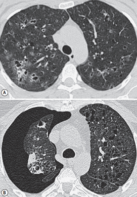
FIGURE 1 ■ Langerhans cell histiocytosis. (A) The characteristic combination of thin-walled cysts and poorly defined nodules, some of which are just beginning to cavitate. (B) Image from a patient with more advanced disease. There are numerous irregularly shaped cysts bilaterally and a pneumothorax on the right.
责任编辑:admin
上一篇:没有了
下一篇:医学文章阅读——Parenchymal Changes

微信公众号搜索“译员”关注我们,每天为您推送翻译理论和技巧,外语学习及翻译招聘信息。