The commonest CT finding of remodelling of bronchi is bronchial wall thickening, which is common in smokers, more particularly those with functional impairment, and more particularly those presenting clinical symptoms of chronic bronchitis. Bronchial wall and lumen irregularities are commonly visualised on CT, reflecting remodelling and deficit in cartilage (Fig. 1). Evidence of moderate tubular (cylindrical) bronchiectasis (most in thelower lobes) is common (29–58%). Bronchiectasis is associated with severe airflow obstruction, more severe COPD exacerbation, lower airway bacterial colonisation, and increased sputum inflammatory markers. Bronchial diverticula, seen as outpouchings in addition to the lumen of mainstem and lobar bronchi, reflect fusion of multiple depressions and dilatations of bronchial gland ducts; these features are best visualised on longitudinal minimal intensity projection thick-slab reconstructions and have been reported in 12% of smokers (mostly in heavy smokers with cough, with severe functional impairment, extensive emphysema and more severe bronchial wall thickening). Bronchomalacia may be demonstrated on expiratory CT as almost complete collapse of the lumen of segmental or subsegmental bronchi, mostly in the lower lobes where the deficit in cartilage is more apparent.
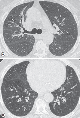
FIGURE 1 ■ COPD patient with airway disease predominant phenotype. Axial CT at the levels of the upper (A) and lower (B) parts of the chest. Few small centrilobular and paraseptal emphysematous spaces in the upper lobes. Bronchial wall thickening, slight bronchial dilatation and lung parenchyma hypoattenuation reflecting obstructive bronchiolitis in the lower lobes.
责任编辑:admin
上一篇:没有了
下一篇:医学文章阅读——Panlobular or Panacinar Emphysema (PLE)

微信公众号搜索“译员”关注我们,每天为您推送翻译理论和技巧,外语学习及翻译招聘信息。