Panlobular or panacinar emphysema is manifested as a generalised decrease of attenuation of the lung parenchyma without focal lucencies. Although this pattern is classically described with α1-antitryspin deficiency, a similar pattern may be seen with severe smoking-related emphysema. The vessels in the affected lung are usually reduced in number and in calibre, straightened and show decreased branching. The appearance of featureless decreased attenuation may sometimes be quite difficult to distinguish from severe obliterative bronchiolitis. The presence of long lines in the lower lobes reflecting fibrosis within the remaining interlobular septa in panlobular emphysema helps distinguish this entity from obliterative bronchiolitis (Fig. 1). Usually these abnormalities are most severe in the lower lobes. As clinical manifestations of PLE associated with α1-protease inhibitor deficiency are often seen in cigarette smokers, focal lucencies due to centrilobular emphysema may be seen in the upper lobes. Occasionally, PLE occurring in smokers is predominant in the upper lungs. Paraseptal emphysema and bullae can also be seen, but are not a major feature of the disease. In severe PLE, the characteristic appearance of extensive lung destruction and the associated paucity of vascular markings may have a diffuse distribution. On the other hand, mild and even moderately severe PLE can be very subtle and difficult to detect radiologically.
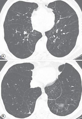
FIGURE 1 ■ Panlobular emphysema in a patient with α1-antitryspin deficiency. Axial CT at the levels of the mild (A) and lower parts (B) of the lung with diffuse lung attenuation and paucity of the pulmonary vessels. The presence of multiple thin lines, particularly throughout the lung bases, reflects a distortion of the anatomical structure of the lung parenchyma and thickening of the remaining interlobular septa by lung fibrosis.
责任编辑:admin
上一篇:没有了
下一篇:医学文章阅读——Emphysema

微信公众号搜索“译员”关注我们,每天为您推送翻译理论和技巧,外语学习及翻译招聘信息。