Strictures of the trachea are usually secondary to damage from a cuffed endotracheal or tracheostomy tube or to external neck trauma. The lesions consist of granulation tissue leading to dense mucosal and submucosal fibrosis associated with distortion of cartilage plates. The two principal sites of stenosis following intubation or tracheostomy are at the level of the stoma or the endotracheal balloon.
On radiography, the stenosis may be seen as a focus of circumferential or eccentric narrowing associated with a segment of increased soft tissue. The size of narrowing is usually well seen at CT and is often concentric. Postintubation stenosis extends for several centimetres and is typically seen above the level of the thoracic inlet. Posttracheostomy stenosis typically begins 1–1.5 cm distal to the inferior stromal margin and extends for 1.5–2.5 cm. Multiplanar reformations accurately determine the site, the length and the degree of the stenosis (Fig. 1). In selected cases, the degree of stenosis may also be attained by use of virtual bronchoscopy
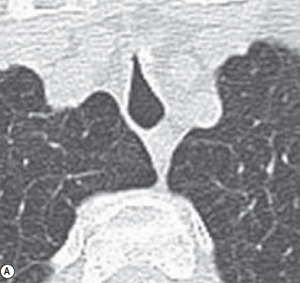
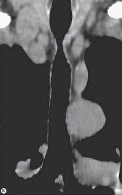
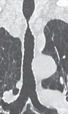
FIGURE 1 ■ Post-intubation tracheal stenosis in a severe COPD patient. (A) Axial CT (lung window). (B) Coronal oblique MPR image (mediastinal window) along the long axis of the trachea. (C) Coronal oblique MPR image (lung window).
责任编辑:admin
上一篇:没有了
下一篇:医学文章阅读——Echinococcosis (Hydatid Disease)

微信公众号搜索“译员”关注我们,每天为您推送翻译理论和技巧,外语学习及翻译招聘信息。