Extensive lymph node calcification is common following tuberculosis and fungal infection, and is occasionally seen with other infections (Fig. 1). It may also be encountered in a variety of other conditions, notably sarcoidosis, silicosis and amyloidosis. Although it may be seen in lymph node metastases from calcifying primary malignancies, such as osteosarcoma, chondrosarcoma and mucinous colorectal and ovarian tumours, lymph node calcification is rare in metastatic neoplasm. It is virtually unknown in untreated lymphoma though it is occasionally seen in nodes involved by Hodgkin's disease following therapy.
CT is the most sensitive technique for the detection of lymph node calcification. Two common patterns of calcification are coarse, irregularly distributed clumps within the node and homogeneous calcification of the whole node. A strikingly foamy appearance is rarely seen with Pneumocystis jiroveci (previously Pneumocystis carinii) infection in AIDS patients and in some cases of metastatic mucinous neoplasms. Sometimes there is a ring of calcification at the periphery of the node—so-called 'eggshell calcification', which is a particular feature of sarcoidosis and of prolonged dust exposure (e.g. mining).
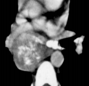
FIGURE 1 ■ High-attenuation lymph nodes. Transaxial image of chest CT shows a calcified mediastinal lymphadenopathy. Such dystrophic calcification is common as a sequela of Histoplasma capsulatum infection; however, it can also be seen with metastatic lymphadenopathy of mucinous adenocarcinomas.
责任编辑:admin
上一篇:没有了
下一篇:医学文章阅读——Non-Seminomatous Germ-Cell Tumours

微信公众号搜索“译员”关注我们,每天为您推送翻译理论和技巧,外语学习及翻译招聘信息。