It is theoretically possible to measure the volume of blood flowing within a vessel using Doppler ultrasound. This requires calculation of the mean blood velocity in the vessel and the cross-sectional area of the vessel. The product of these two values is the volume of blood flowing. In practice, there are significant uncertainties about both values, which may lead to errors in excess of 100%. Calculation of the mean velocity should take into consideration the distribution of velocities across the vessel in each time interval and the average of these over several cardiac cycles (Fig. 1). It is also assumed that the vessel is uniformly insonated and that signals are detected from all the blood in the cross-section of the vessel. In practice, slow-moving blood is missed and the range gate or beam width may encompass only a fraction of the vessel's area.
These errors may not be obvious from visual inspection of the spectral display. If the crosssectional area of a vessel is calculated from a single diameter, any error is squared. Measuring the area from an image at right angles to the vessel axis may be more accurate, but this information cannot be obtained at the same time and site as the Doppler signal, owing to the inappropriate beam/vessel angle. Volume blood flow measurements in the carotid and femoral vessels and in the abdomen have not been shown to be of clinical value.
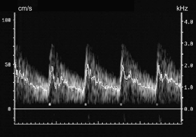
FIGURE 1 ■ Calculation of time-averaged mean velocity. The weighted mean velocity during each time interval has been calculated. The average of these throughout the trace is a close estimate of the true mean velocity.
责任编辑:admin
上一篇:没有了
下一篇:医学文章阅读——Echogenicity

微信公众号搜索“译员”关注我们,每天为您推送翻译理论和技巧,外语学习及翻译招聘信息。