Silica causes three distinct clinical patterns of lung disease which are related to both level and duration of exposure. The earliest radiographic changes of silicosis and coal worker's pneumoconiosis (CWP) are nearly identical. Typical appearances are a profusion of small (1–3 mm) round nodules distributed in the posterior aspects of the upper two-thirds of the lung. Radiologically, the only difference between simple CWP and simple silicosis is that the nodules in CWP are often smaller. With advancing disease, the nodules increase in size and number to involve all lung zones. The nodules are sometimes calcified. Hilar and mediastinal lymph node enlargement with calcification of the eggshell type is not uncommon and may be seen on the chest radiograph or CT. On CT, the micronodules are sharply defined and distributed throughout the lungs but are frequently most numerous in the upper lung zones. The nodules may be centrilobular or subpleural in location; the subpleural micronodules may become confluent, forming a 'pseudo-plaque'.
Progressive massive fibrosis (PMF) refers to the coalescence of large nodules and is much more common in silicosis than in CWP. On the chest radiograph, PMF is seen as mass-like opacities, typically in the posterior upper lobes and associated with contraction of the upper lobes and hilar elevation. Sequential evaluation of these masses often demonstrates migration towards the hila, leaving a peripheral rim of cicatricial emphysema. The outer margins of PMF often parallel the contour of the adjacent chest wall. CT confirms the architectural distortion associated with PMF (Fig. 1). Large lesions (>5 cm) often show irregular low-attenuation regions on CT indicative of necrosis. Frank cavitation is a less frequent finding and when present should always raise the suspicion of tuberculosis (conventional or atypical). Unilateral or asymmetric PMF may be distinguished from lung cancer by the presence of lobar volume loss and peripheral emphysema.
Classic HRCT Findings
• Well-defined dense centrilobular and subpleural nodules, concentrated in the upper zones posteriorly
• Hilar and mediastinal lymph node enlargement, with or without calcification, is frequent
• Upper lobe irregular masses characterise progressive massive fibrosis
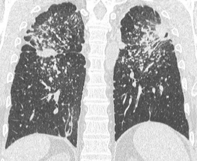
FIGURE 1 ■ Progressive massive fibrosis in coalworker's pneumoconiosis. Mass-like opacities are seen bilaterally in the upper lobes in association with multiple small nodules and calcified mediastinal lymphadenopathy.
责任编辑:admin
上一篇:没有了
下一篇:医学文章阅读——The International Labour Office Classification

微信公众号搜索“译员”关注我们,每天为您推送翻译理论和技巧,外语学习及翻译招聘信息。