Non-specific interstitial pneumonia (NSIP) is characterised by varying degrees of interstitial inflammation and fibrosis without the specific features that allow a diagnosis of UIP. While NSIP may have significant fibrosis, it is usually temporally uniform (in comparison to UIP), and fibroblastic foci and honeycombing are absent or scanty. Although the clinical features of idiopathic NSIP resemble those of UIP, prognosis is considerably better. Non-idiopathic NSIP is most often found on lung biopsy in patients with connective tissue disease and may be the predominant histopathological pattern in some cases of drug-induced lung disease and chronic hypersensitivity pneumonitis. On HRCT, ground glass with or without associated distortion of airways is usually the dominant pattern (Fig. 1). Reticular abnormalities are common, but honeycombing is sparse or absent even when other signs of fibrotic changes are evident. Abnormalities are usually peribronchovascular or peripheral, although they may sometimes spare the subpleural lung. In general, NSIP may be distinguished from UIP on CT by a more prominent component of ground-glass attenuation and a finer reticular pattern in the absence of honeycombing. However, the variability of CT appearances reflects the heterogeneity of the pathological processes encompassed by NSIP and a confident diagnosis of NSIP based on CT alone is less readily made than in cases of UIP. Consolidation is reportedly a highly variable feature (0–98%) and this discrepancy probably reflects the fact that some patients with non-idiopathic NSIP have significant amounts of histological organising pneumonia, making classification of individual cases difficult.
Classic HRCT Findings
• Bilateral ground-glass opacities and superimposed fine reticulation
• Predominantly peripheral, but also patchy or band-like
• Honeycomb pattern minimal or absent
• Traction bronchiectasis and bronchiolectasis
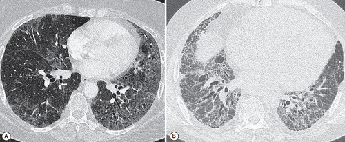
FIGURE 1 ■ Non-specific interstitial pneumonia. (A) The predominant abnormality is patchy, bilateral ground-glass opacification, mild reticulation and traction bronchiectasis. (B) In this fibrotic form, reticulation is more extensive and traction bronchiectasis more severe. However, there is no frank honeycombing destruction.
责任编辑:admin
上一篇:没有了
下一篇:医学文章阅读——Mosaic Attenuation Pattern Of Diffuse Lung Disease

微信公众号搜索“译员”关注我们,每天为您推送翻译理论和技巧,外语学习及翻译招聘信息。