Although the aetiology of pleural thickening (or a combination of thickening and pleural effusion) will be determined in most patients by a combination of clinical history and imaging features making biopsy unnecessary, some patients require further investigation and histological confirmation (Fig. 1). This may either be by percutaneous biopsy or under direct visualisation at medical or video-assisted thoracoscopy. The use of closed needle pleural biopsy such as the Abram's biopsy has almost disappeared in all areas other than those endemic for tuberculosis, following evidence comparing its efficacy to image-guided cutting needle biopsy, particularly in patients with suspected pleural malignancy. Whilst percutaneous biopsy has been shown to have a sensitivity of almost 90% for mesothelioma and for other pleural malignancies such as adenocarcinoma, it is unable to provide a tissue diagnosis and treat an associated pleural effusion in a single procedure. By comparison, medical thoracoscopy, which has become the technique of choice for patients with suspected malignant effusions, and percutaneous image-guided biopsy (mostly for patients unsuitable for thoracoscopy) allow combined diagnosis and therapy in a single procedure. In patients with suspected mesothelioma, a tissue diagnosis should be achieved using as few interventions as possible, because of the known incidence of biopsy and drain track tumour seeding (Fig. 2).
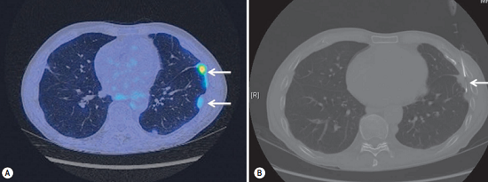
FIGURE 1 ■ (A) PET-CT performed in a patient with suspected malignant pleural thickening, but the initial percutaneous biopsy was negative. Note how the two large left-sided pleural nodules, arrowed, have differing 18F-fluorodeoxyglucose avidity. (B) The more avid of the two nodules has been targeted for biopsy, arrowed.
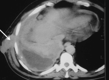
FIGURE 2 ■ CT, demonstrating mesothelioma growing, arrowed, along the site of a prior pleural drain.
责任编辑:admin
上一篇:没有了
下一篇:医学文章阅读——Chest Drains

微信公众号搜索“译员”关注我们,每天为您推送翻译理论和技巧,外语学习及翻译招聘信息。