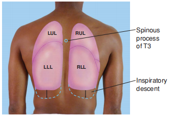
Picture the lungs and their fissures and lobes on the chest wall. Anteriorly, the apex of each lung rises approximately 2 to 4 cm above the inner third of the clavicle (Fig. 1). The lower border of the lung crosses the 6th rib at the midclavicular line and the 8th rib at the midaxillary line. Posteriorly, the lower border of the lung lies at about the level of the T10 spinous process (Fig. 2). On inspiration, it descends in the chest cavity during contraction and descent of the diaphragm.

FIGURE 1. The anterior lungs.

FIGURE 2. The posterior lungs.
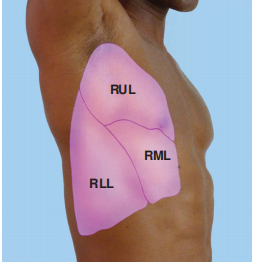
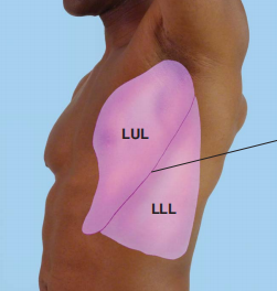
Each lung is divided roughly in half by an oblique (major) fissure. This fissure may be approximated by a string that runs from the T3 spinous process obliquely down and around the chest to the 6th rib at the midclavicular line (Fig. 3). The right lung is further divided by the horizontal (minor) fissure. Anteriorly, this fissure runs close to the 4th rib and meets the oblique fissure in the midaxillary line near the 5th rib. The right lung is thus divided into upper, middle, and lower lobes (RUL, RML, and RLL). The left lung has only two lobes, upper and lower (LUL, LLL) (Fig. 4). Each lung receives deoxygenated blood from its pulmonary artery. Oxygenated blood returns from each lung to the left atrium via the pulmonary veins.

FIGURE 3. Right lung lobes and fissures.

FIGURE 4. Left lung lobes and fissures.
责任编辑:admin
上一篇:没有了
下一篇:医学文章阅读——Steps for Using the Ophthalmoscope

微信公众号搜索“译员”关注我们,每天为您推送翻译理论和技巧,外语学习及翻译招聘信息。