Histoplasma capsulatum is a fungus found in moist soil and in bird or bat excreta in many parts of the world but human infection is endemic in areas such Ohio–Mississippi and St. Lawrence River valleys (North America).
Though nodules greater than 3 cm may be seen (Fig.1), the most common radiographic findings consist of diffuse nodular opacities of 3 mm or less in diameter, nodules greater than 3 mm in diameter, small linear opacities and focal or patchy areas of consolidation. As these findings are not seen on chest radiography in approximately 40% of patients with pulmonary histoplasmosis, CT is requested as a more sensitive imaging investigation. Hilar and mediastinal lymph nodes are frequently enlarged. Chronic pulmonary histoplasmosis radiologically resembles post-primary tuberculosis. In some cases, fibrosing mediastinitis may develop and can lead to constriction of mediastinal structures, including the airways, superior vena cava, pulmonary arteries and pulmonary veins.
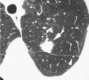
FIGURE 1 ■ Histoplasmoma. A 62-year-old asymptomatic woman living in an area endemic for histoplasmosis. An incidental nodule was found on routine chest radiography. CT shows a rounded opacity in the left upper lobe (arrow). A 3-mm nodule (arrowhead) is also seen in the superior segment of the left lower lobe.
责任编辑:admin
上一篇:没有了
下一篇:医学文章阅读——CHANGING SPECTRUM OF HIV INFECTIONS: 30 YEARS LATER

微信公众号搜索“译员”关注我们,每天为您推送翻译理论和技巧,外语学习及翻译招聘信息。