A clinical diagnosis of pneumonia is usually established on the basis of clinical symptoms, laboratory findings and chest radiography. Though different patterns of pneumonia are associated with certain underlying microorganisms, it has to be clearly stated that there is no specific radiological pattern of pneumonia caused by one particular microorganism. Overlap of imaging findings also with respect to course over time makes the differentiation of aetiologies based solely on the radiograph unreliable, and distinguishing pneumonia from conditions such as left heart failure and pulmonary embolism may sometimes be difficult, especially as patients with pre-existing lung disease (severe emphysema, interstitial lung disease, etc.) who may develop very atypical patterns of pneumonia.
New emerging pathogens have been recognised such as community-acquired methicillin-resistant S. aureus, human metapneumovirus, avian influenza A viruses (H5N1), coronavirus associated with severe acute respiratory syndrome (SARS) and swine flu (H1N1).
Chest radiography remains an important component of the evaluation of a patient with a suspicion of pneumonia, and usually is the first examination to be obtained. CT, preferably with thin (<2 mm thick) slices, has been shown to be more sensitive than radiography in the detection of subtle abnormalities and may show findings suggestive of pneumonia up to 5 days earlier than chest radiographs. CT is recommended in patients with clinical suspicion of infection and normal or non-specific radiographic findings (Fig. 12-1) and in patients with increased risk of pulmonary infection (e.g. neutropenia). CT is also indicated in patients with pneumonia and persistent or recurrent pulmonary opacities to diagnose or rule out underlying or alternative disease processes.
The presence of a radiographically visibile opacification is part of the definition of pneumonia, according to the American Thoracic Society (ATS), though there might be a time delay of several hours between onset of clinical symptoms and radiographic changes, and specific conditions may further the delay or cause a negative chest radiograph.
Regression of pneumonia over time varies with the underlying organism, patient comorbidity and patient age and can take between 1 and 2 weeks or up to 2 months.
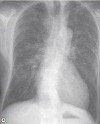
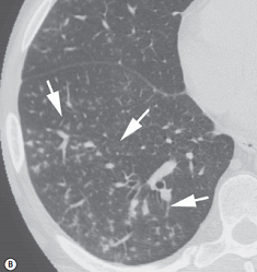
FIGURE 1 ■ Cellular bronchiolitis. A 71-year-old man with fever of 48 h duration. (A) Posteroanterior chest radiograph is normal. (B) Complementary CT shows centrilobular branching nodular and linear opacities resulting in a 'tree-in-bud' appearance (arrows). Mycoplasma bronchiolitis was diagnosed.
责任编辑:admin
上一篇:没有了
下一篇:医学文章阅读——Constrictive Pericarditis

微信公众号搜索“译员”关注我们,每天为您推送翻译理论和技巧,外语学习及翻译招聘信息。