Inflammation of the pericardium (pericarditis) may occur in response to a variety of insults. Viral infection is the most common cause of pericarditis in the United States, the most common agents being Coxsackie group B and echoviruses. Pericarditis typically results in cellular proliferation, or the production of fluid (pericardial effusion) or fibrin, either alone or in combination. Thickening of the pericardium occurs because of fibrinous exudates and oedema. Causes include myocardial infarction (acute or post-myocardial infarction referred to as Dressler's syndrome), pericardiotomy, mediastinal irradiation, infection (viral or bacterial), connective tissue disease (rheumatoid arthritis, systemic lupus erythematosus), metabolic disorders (uraemia, hypothyroidism), neoplasia and AIDS.
The most common imaging manifestation of acute pericarditis is a pericardial effusion, the nature of the fluid varying with the underlying cause. Thickened inflamed pericardium can appear as moderate-to-high signal intensity on spin-echo MRI, and pericardial enhancement may be seen on either MRI or CT performed after intravenous contrast medium administration. Delayed images on contrast medium-enhanced CT are useful for demonstrating pericardial enhancement (Fig. 1).
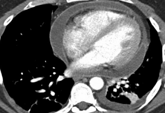
FIGURE 1 ■ Pericarditis. A 30-year-old man with new-onset of left-sided chest pain. Axial contrast medium-enhanced CT shows diffuse pericardial thickening and a moderate-sized pericardial effusion. This was caused by viral pericarditis.
责任编辑:admin
上一篇:没有了
下一篇:医学文章阅读——Cardiac Tamponade

微信公众号搜索“译员”关注我们,每天为您推送翻译理论和技巧,外语学习及翻译招聘信息。