Fatty tumours of the mediastinum are rare. On chest radiography, regardless of whether they are benign or malignant, fatty tumours are seen as well-defined round or oval mediastinal masses.
Mediastinal lipomas constitute 2% of all mediastinal tumours. They can occur in any part of the mediastinum but are most common in the prevascular space. Benign lipomas are soft and do not compress surrounding structures unless they are very large. On CT they show uniform fat attenuation. Their boundaries are smooth and sharply demarcated from adjacent mediastinal structures.
Mediastinal liposarcomas are rare malignant fat-containing tumours. They may occur anywhere in the mediastinum. In contradistinction to benign lipomas, they usually contain large areas of soft-tissue density material. Histological differentiation between lipoma and liposarcoma depends on the presence of mitotic activity, cellular atypia, neovascularisation and tumour infiltration.
CT findings include heterogeneous attenuation with significant soft tissue within a mass with fat attenuation, poor definition of adjacent mediastinal structures and infiltration or invasion of mediastinal structures (Fig. 1).
Lipoblastoma, a benign tumour of childhood, contains fat and soft tissue. Occasionally, the amount of fat attenuation is relatively small. CT findings are similar to liposarcomas.
Angiomyolipoma and myelolipoma are both benign tumours which may show a combination of soft-tissue and fat attenuation on CT and therefore can be indistinguishable from liposarcoma on imaging. Angiomyolipomas and myelolipomas are rare in the mediastinum.
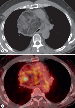
FIGURE 1 ■ Liposarcoma. A 58-year-old man with gradually worsening dyspnoea was found to have a large anterior mediastinal mass when evaluated by chest CT. This mass has a predominantly fat attenuation with internal thick septations and mural nodules and exerts significant mass effect upon the airway (A). PET-CT shows significant FDG uptake of the soft-tissue components of the mass (B).
责任编辑:admin
上一篇:没有了
下一篇:医学文章阅读——Sympathetic Ganglion Tumours

微信公众号搜索“译员”关注我们,每天为您推送翻译理论和技巧,外语学习及翻译招聘信息。