Peripheral nerve tumours are the most common mediastinal neurogenic tumours. They typically originate in an intercostal nerve in the paravertebral region. Most are benign. Radiologically, the benign tumours (neurofibromas and schwannomas) present as well-defined round or oval posterior mediastinal masses. Pressure deformity causing a smooth, scalloped indentation on the adjacent ribs, vertebral bodies, pedicles or transverse processes is common, particularly with larger lesions.
The scalloped cortex is usually preserved and is often thickened. These bone changes are diagnostic of a neurogenic lesion, the only differential diagnosis being that of a lateral thoracic meningocele. The rib spaces and the intervertebral foramina may be widened by the tumour. On CT the tumours may be homogeneous or heterogeneous, usually enhancing heterogeneously. Punctate foci of calcification may be seen. Care must be taken on CT, however, as these lesions are often homogeneous and low in attenuation (from the myelin content). The net effect is a lesion that can mimic a duplication cyst. As a general rule, a posterior mediastinal lesion should not be called a cyst unless there is a clear vertebral anomaly or communication with the spinal canal.
On MR, neurofibromas and schwannomas have low-to-intermediate signal intensity on T1-weighted images and may have characteristic high signal intensity peripherally and low signal intensity centrally (target sign) on 2-weighted images; and enhance after gadolinium. Ten percent of paravertebral neurofibroma extend into the spinal canal and appear as dumbbell-shaped masses with widening of the affected neural foramen.
Malignant tumours of nerve sheath origin (Fig. 1) are rare spindle cell sarcomas, typically occurring in the third to fifth decades, although they may occur earlier in patients with neurofibromatosis type 1. Radiologically, the masses are usually larger than 5 cm in diameter. Although MR cannot reliably differentiate benign from malignant neurogenic tumours, sudden change in size of a pre-existing mass, the development of heterogeneous signal intensity (caused by haemorrhage and necrosis) or infiltration of adjacent mediastinum or chest wall is cause for concern. Haematogenous metastases to the lung have been reported but lymph node metastasis is rare.
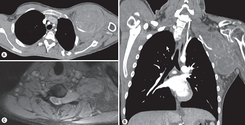
FIGURE 1 ■ Malignant nerve sheath tumour. A 23-year-old with left axillary mass and left shoulder pain. (A) Axial and (B) coronal contrast medium-enhanced CT images show a large heterogeneously enhancing mass in the left axilla, which encases the left subclavian artery. Axial contrast medium-enhanced MR image demonstrates that this enhancing mass expands the neural foramen of the spine, with no erosion of the vertebral body, suggesting that this is a neurogenic tumour (C).
责任编辑:admin
上一篇:没有了
下一篇:医学文章阅读——Lymph Node Enlargement

微信公众号搜索“译员”关注我们,每天为您推送翻译理论和技巧,外语学习及翻译招聘信息。